Your trip to the dentist in Airdrie should include regularly scheduled dental x-rays. An x-ray is an imaging test that dentists use to get a clear image of your teeth and jaw to be able to plan what course of treatment to follow. This may mean filling cavities, dental braces or just a regular pat on the back for good oral hygiene.
When having x-rays, various parts of your body absorb radiation at different rates. The calcium in your bones draws the most amount of radiation. As a result, your bones look white after the imaging. Fat, muscles and other soft tissues absorb less, which is why they look grey in the image.
Of course, a dental radiograph is not a one-size-fits-all diagnostic procedure. Doctors and specialists use x-rays to determine any possible oral care issues, such as impacted or abnormal development of the teeth, wisdom tooth and even gum disease. They’re divided into two main categories: intraoral and extraoral.
Intraoral Radiographs
In this type of procedure, the x-ray film is placed inside the mouth. This type of radiograph is the most common among all the types of dental x-rays. Intraoral radiographs provide a tremendous amount of detail, letting your dentist find cavities and check the health of your entire tooth–enamel to root. Types of intraoral x-rays include:
- Bite-wing X-rays unravel the upper and lower teeth in a specific area in your mouth. This type of radiograph shows the molars (backmost teeth) and the premolars. After biting a wing-shaped device, one or more images are taken. Your dentist checks the radiographic image for any signs of decay and gum disease. This type of imaging can also help your dentist during restorations and fillings.
- Periapical X-rays are captured almost the same way as bite-wing x-rays, but only one tooth is examined. This type of x-ray shows the entire tooth from the crown down to the root to check changes around the tooth and neighbouring bone structures.
- Occlusal X-rays show nearly the entire arch of the teeth on either the upper or lower jaw.
Extraoral Radiographs
This kind of procedure provides information on the jaw and skull. Unlike intraoral x-rays, this type of radiograph gives fewer details of an individual tooth. Here are examples and brief explanations of each:
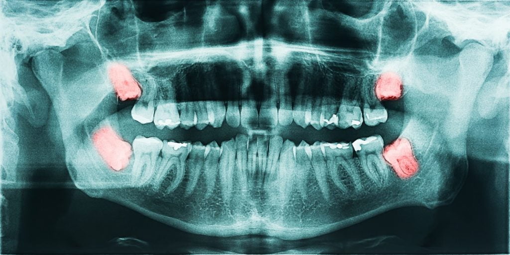
- Panoramic X-rays show the entire mouth area to detect developing and impacted teeth in the upper or lower jaw. Some dentists also use this procedure to help diagnose tumours.
- Tomograms examine structures in the mouth that are difficult to see because of obstacles and nearby structures blocking the typical view. Only one layer is shown while the rest are blurred out.
- Cephalometric projections show an entire side of the head to help orthodontists determine the best teeth-realignment approach.
- Dental Computed (CT) Tomography looks at interior structures in 3D to detect problems in facial bones such as cysts, tumours, and fractures.
- Cone Beam CT creates high-quality 3-D images of your soft tissues, nerves, and bones to help dentists in tooth implant procedures
- Magnetic Resonance Imaging (MRI) takes a 3-D view of the oral cavity including your jaw and teeth to evaluate soft tissues
Final Thoughts
This list is not exhaustive, other types of specialized intraoral and extraoral radiographs are not mentioned. Both types of x-rays can be vital in helping your dentists create a comprehensive dental care plan and treatment specifically tailored for you. Hopefully, this gives you a clear picture (pun intended) of how important x-rays are in making your Airdrie dentist’s work more straightforward, and your smile brighter.
Rest assured that at Airdrie Springs Dental, we only use digital X-rays which produce the lowest possible dose of radiation